
Uveal Tract Anatomy & Physiology
Curriculum lesson · updated 2026-07-30
Pending review from section expertOn this page
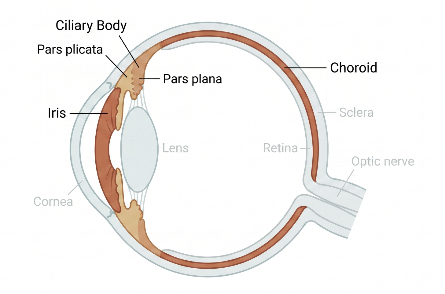
The uvea is the eye's middle, pigmented, highly vascular layer between the outer fibrous coat (sclera/cornea) and the inner neurosensory retina, composed of the iris, ciliary body, and choroid; inflammation of any part is called uveitis.1 The iris is the most anterior uveal structure, a pigmented diaphragm controlling pupil size via sphincter and dilator muscles. The ciliary body, just posterior to the iris root, has two functional parts: the pars plicata (produces aqueous humor, anchors lens zonules) and the pars plana (smooth posterior extension serving as the vitreous humor's attachment point). The choroid is the most posterior and extensive uveal structure, densely vascular, supplying the outer retina including photoreceptors and RPE. This subdivision directly defines uveitis classification: anterior uveitis affects the iris/ciliary body (41-60% of cases), intermediate uveitis affects the pars plana/peripheral retina (9-15%), posterior uveitis involves the choroid/retina (17-23%), and panuveitis involves all uveal layers (7-32%).1
The eye, including the uveal tract, maintains immune privilege — tightly regulated tolerance limiting vision-threatening inflammation from minor antigenic challenge. This privilege is maintained through anatomical barriers (blood-ocular barriers), local immunomodulatory mediators, regulatory immune-cell interactions, and anterior chamber-associated immune deviation (ACAID), a form of systemic tolerance induced by intraocular antigen exposure.2 Ocular immune privilege is best understood as dynamic and conditional rather than absolute: across autoimmune uveitis, corneal transplantation, AMD, diabetic retinopathy, ocular tumors, and allergic conjunctivitis, disease is generally associated with partial disruption, remodeling, or exploitation of these mechanisms rather than complete loss.2 These alterations vary by ocular compartment but recurrently involve disrupted immune homeostasis, increased inflammatory signaling, altered leukocyte activity, impaired barrier integrity, and reduced local immune restraint.2
Untreated uveitis, regardless of anatomic subtype, can cause cataract, glaucoma, macular edema, retinal detachment, and optic nerve damage, reflecting the uvea's direct anatomic and vascular connections to the lens, angle, retina, and optic nerve.1 Uveitis predominantly affects adults 20-50 years old; while a substantial proportion is idiopathic (27-51% in the US/Europe), 37-49% is associated with an identifiable systemic disease such as axial spondyloarthritis, underscoring why uveitis often warrants a systemic, not just ocular, workup.1 Recognizing which elements of immune privilege are preserved or disrupted in a given setting helps explain why inflammation, transplantation, tumors, and allergy behave differently across ocular tissues, and may support more precise immune-directed treatment.2
- Uveitis anatomic classification (anterior, intermediate, posterior, panuveitis) directly maps onto which uveal structure is inflamed1
- The pars plana, not the pars plicata, is the anatomic landmark defining intermediate uveitis and the site of vitreous base attachment1
- Ocular immune privilege is a dynamic, conditional system, not an all-or-nothing state, maintained by anatomical barriers, local mediators, and ACAID-mediated tolerance2
- A substantial proportion of uveitis is linked to systemic disease, making a directed systemic review essential1
- Common pitfall: treating "uveitis" as a single disease rather than a group of conditions defined by which anatomic compartment is involved1
- Common pitfall: confusing the pars plicata (aqueous production, zonules) with the pars plana (vitreous attachment, intermediate uveitis landmark)
- Common pitfall: assuming ocular immune privilege is either fully intact or completely absent rather than a graded, context-dependent state2
Uveal Tract Anatomy & the Uveitis Classification It Defines
Clinical Skills for This Topic
Slit-lamp grading of anterior chamber cell and flare →
Localize and grade inflammation in the anterior uveal compartment.
Performing a dilated posterior segment exam in uveitis →
Evaluate the pars plana, choroid, and retina to determine intermediate, posterior, or panuveitis involvement.
Screening for associated systemic disease
Recognize when uveitis anatomic pattern and history warrant referral for conditions like axial spondyloarthritis or sarcoidosis.
Practice Questions
Question 1 of 6
Score: 0/0
What three structures make up the uveal tract?
Saved in this browser.