
Retina Anatomy & Physiology
Curriculum lesson · updated 2026-07-30
Pending review from section expertOn this page
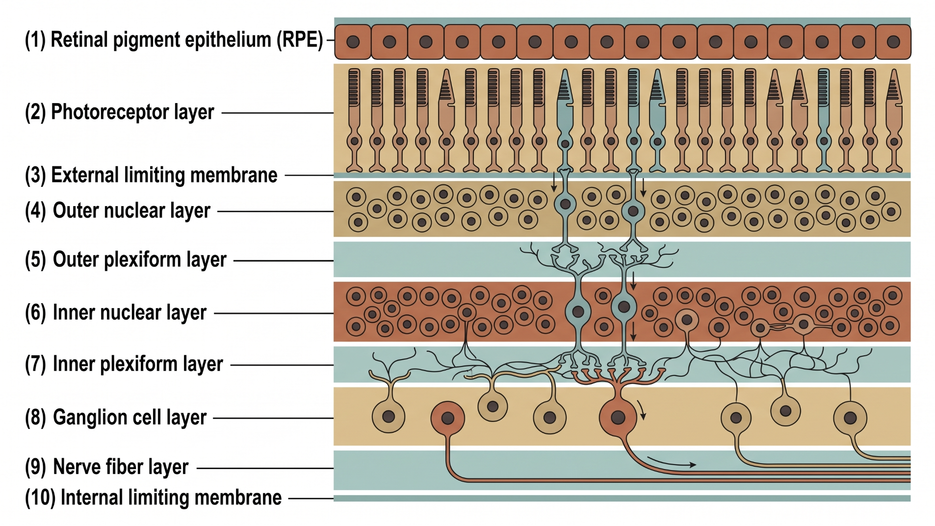
The retina is a thin, ten-layered neurosensory tissue lining the posterior segment that converts light into electrical signals transmitted via the optic nerve to the visual cortex. From the vitreous side inward: internal limiting membrane, nerve fiber layer, ganglion cell layer, inner plexiform layer, inner nuclear layer, outer plexiform layer, outer nuclear layer, external limiting membrane, and photoreceptor inner/outer segments. The retinal pigment epithelium (RPE) forms the outermost layer, adjacent to Bruch's membrane and the choriocapillaris of the choroid, and is indispensable to photoreceptor survival despite not being light-sensitive itself.1 The macula is specialized for high-acuity central vision; its center, the fovea, is cone-dense and thinner than surrounding retina because inner layers are displaced laterally to minimize light scatter before reaching the photoreceptors.
The RPE performs a remarkable range of functions for a single cell layer. It forms the outer blood-retina barrier through tight junctions between adjacent cells, which — together with the inner blood-retina barrier at the retinal vasculature — regulates what reaches the outer retina and helps create an immune-privileged space in the eye.1 RPE cells absorb stray light focused onto the retina by the lens, continuously phagocytose shed photoreceptor outer segment discs, and perform spatial buffering of potassium in the subretinal space in response to changing illumination.1 RPE cells are long-lived, essentially post-mitotic cells that must sustain these functions for decades under continuous oxidative, metabolic, and phagocytic stress.2
A central RPE function is the visual (retinoid) cycle: after a photoreceptor's visual pigment absorbs light and its chromophore isomerizes, the RPE re-isomerizes the photopigment back to 11-cis retinal, regenerating rhodopsin for the next phototransduction cycle.1 The RPE also removes water from retinal tissue and secretes factors maintaining structural stability of both the choriocapillaris and photoreceptors.1 Because these barrier and secretory functions maintain an immune-privileged outer retina, loss or alteration of RPE function can directly impair vision independent of any primary photoreceptor defect.1
Because RPE cells are long-lived and largely non-dividing, age-related change accumulates gradually across several interacting domains: structural remodeling, mitochondrial/metabolic imbalance, incomplete degradative clearance, and chronic low-grade inflammatory signaling.2 Rather than widespread cell loss, RPE aging is better described as progressive decline in cellular organization, buffering capacity, and functional precision — a gradual loss of coordination among homeostatic systems rather than abrupt failure of a single pathway.2 This decline increases vulnerability to age-related retinal dysfunction, though RPE aging should not be understood simply as an early, preclinical stage of age-related macular degeneration — it is a broader phenomenon relevant to aging in long-lived, post-mitotic tissue generally.2
- RPE barrier or transport failure disrupts the outer blood-retina barrier, contributing to subretinal fluid accumulation seen in exudative retinal disease
- Impaired RPE phagocytosis of outer segments or visual cycle dysfunction underlies several inherited retinal dystrophies
- Age-related decline in RPE homeostatic capacity is a contributing framework for understanding age-related macular degeneration, though RPE aging and AMD are not equivalent processes2
- The fovea's thinned, displaced inner retinal layers explain why central vision is exquisitely sensitive to even small anatomic disruption at the macula
- The RPE is not light-sensitive itself, but photoreceptor survival depends entirely on its barrier, transport, phagocytic, and visual-cycle functions1
- The outer blood-retina barrier is formed by RPE tight junctions, distinct from the inner blood-retina barrier formed by the retinal vasculature
- RPE cells are long-lived and largely non-dividing, making them vulnerable to cumulative, decades-long stress rather than acute cell loss2
- The visual cycle's re-isomerization of 11-cis retinal in the RPE is required to regenerate visual pigment after every photobleaching event1
- Common pitfall: treating the RPE as a passive pigmented backdrop rather than an actively functioning barrier, transport, and metabolic tissue1
- Common pitfall: equating normal age-related RPE change with age-related macular degeneration, rather than recognizing AMD as a distinct disease process2
- Common pitfall: attributing all photoreceptor dysfunction to the photoreceptors themselves without considering RPE-level failure
Retinal Layers & the RPE's Supporting Roles
Clinical Skills for This Topic
Dilated fundus examination of the retina and macula →
Assess retinal layers, macular architecture, and pigmentary changes reflecting RPE health.
Recognizing subretinal versus intraretinal fluid patterns
Localize pathology to the RPE/outer blood-retina barrier versus the retinal vasculature/inner blood-retina barrier.
Correlating central acuity loss with foveal anatomy
Understand why even small anatomic disruption at the fovea produces disproportionate central visual loss.
Practice Questions
Question 1 of 6
Score: 0/0
What is the retinal pigment epithelium's relationship to light sensitivity?
Saved in this browser.
References
- [1]Strauß O, Wildner G. Structure and function of the retinal pigment epithelium. Handbook of Clinical Neurology. 2026.
- [2]Yang Y, Liu P, Li J, Deng Y, Xiao L, Peng Q, Peng J. Retinal Pigment Epithelium Ageing: Cellular and Molecular Mechanisms of Long-Term Homeostasis and Age-Related Dysfunction. Cells. 2026.