
Pterygium
Curriculum lesson · updated 2026-07-29
Pending review from section expertOn this page
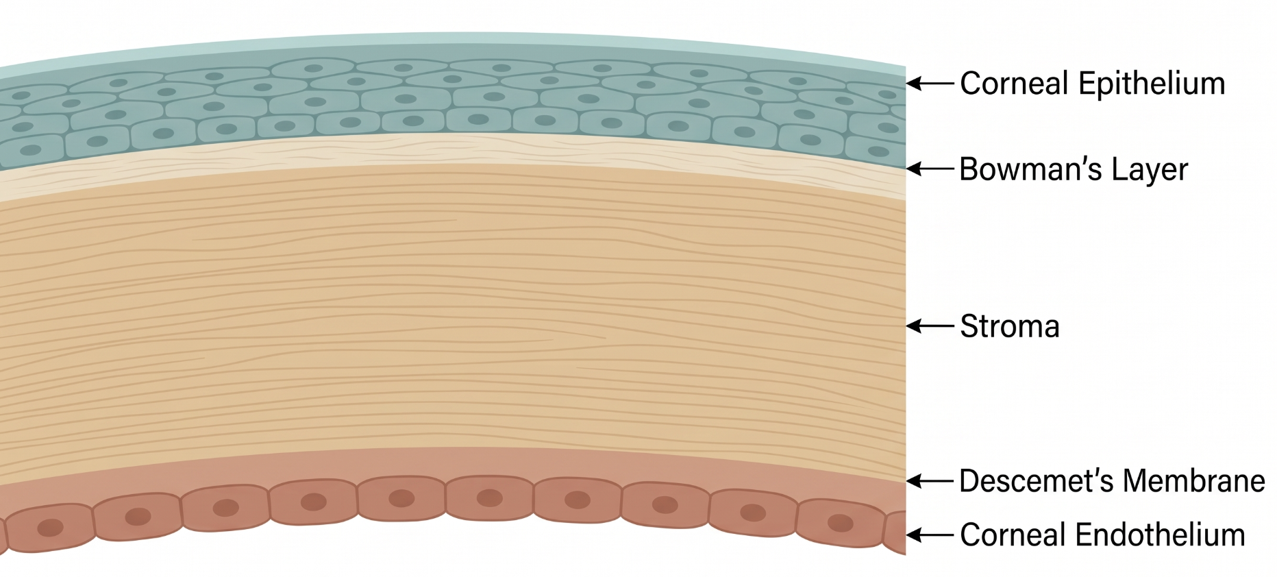
The limbus — the transition zone between the clear cornea and the vascularized, opaque conjunctiva/sclera — is a critical anatomical boundary. It houses limbal stem cells responsible for continuously regenerating corneal epithelium and normally acts as a barrier preventing conjunctival tissue from encroaching onto the cornea. Pterygium represents a breakdown of this boundary: a wing-shaped fibrovascular growth of degenerated conjunctival tissue that crosses the limbus and extends onto the corneal surface, typically nasally.1
Pterygium pathogenesis is complex and incompletely understood, but chronic ultraviolet (UV) light exposure is the most strongly and consistently implicated environmental trigger.1 Proposed mechanisms include UV-induced limbal stem cell alterations, local inflammation, and abnormal fibrovascular tissue remodeling. Molecular research has implicated interactions between the tumor suppressor p53 and its regulator MDM2 in pterygium tissue, an area of ongoing investigation that may inform future, less toxic adjuvant treatments.1
Many pterygia are asymptomatic or minimally symptomatic, noted incidentally or as a visible growth on the white of the eye. Symptomatic patients report foreign-body sensation, mild chronic redness, and cosmetic concern. As the lesion grows toward the visual axis, it can induce irregular astigmatism and eventually direct visual axis obstruction — both indications to consider surgery.
Differential diagnosis includes pinguecula (similar degeneration but does not cross the limbus onto the cornea), conjunctival intraepithelial neoplasia or squamous cell carcinoma (can mimic an atypical or rapidly changing pterygium — always consider with atypical vascularity or gelatinous appearance), and pseudopterygium (a conjunctival adhesion to the cornea following prior inflammation, trauma, or surgery, rather than a primary degenerative growth).
Diagnosis is clinical, based on the characteristic triangular fibrovascular growth with its apex pointing toward the corneal center. Corneal topography can help quantify induced astigmatism when the lesion is enlarging or approaching the visual axis. Biopsy with histopathology is reserved for atypical-appearing lesions to exclude conjunctival neoplasia.
Most pterygia are managed conservatively with observation, UV-protective sunglasses, and lubricating drops. Surgical excision is considered for lesions that are visually significant, rapidly growing, causing persistent irritation unresponsive to conservative measures, or of uncertain diagnosis.
Excision with conjunctival autografting remains preferred over bare sclera excision to reduce recurrence.1 Adjuvant therapies (topical cyclosporine, mitomycin-C, 5-fluorouracil, beta-irradiation) can further reduce recurrence but carry their own toxicity risk and are used selectively.1 A meta-analysis comparing modified sutureless glue-free autograft fixation (using the patient's own blood) versus conventional sutures found the sutureless approach had a shorter operation time and less foreign-body sensation, but a higher risk of graft dehiscence and retraction, with no significant difference in overall recurrence between techniques.2
- Not all conjunctival growths near the limbus are pterygia — always consider conjunctival intraepithelial neoplasia in atypical-appearing or rapidly changing lesions
- Sutureless glue-free grafting is faster and more comfortable postoperatively but carries a higher risk of graft dehiscence/retraction than sutured fixation, with similar overall recurrence
- Conjunctival granuloma is a recognized, and in some series more common, postoperative complication than recurrence itself — counsel patients accordingly
- Common pitfall: assuming every triangular conjunctival-corneal lesion is a benign pterygium without considering neoplastic mimics
- Common pitfall: recommending surgery for all pterygia regardless of visual/symptomatic significance
Pterygium: When to Watch vs. Operate
Clinical Skills for This Topic
Slit-lamp assessment of pterygium extent and activity →
Assess how close the lesion is to the visual axis, look for Stocker's line, and judge vascularity/inflammation to distinguish active from quiescent lesions.
Corneal topography to quantify induced astigmatism
Useful when a growing pterygium is causing irregular astigmatism, to help decide whether surgical referral is warranted.
Recognizing atypical features warranting biopsy
Learn to distinguish a typical pterygium from atypical vascularity, gelatinous appearance, or rapid change that should prompt biopsy to exclude conjunctival neoplasia.
Practice Questions
Question 1 of 6
Score: 0/0
What environmental factor is most strongly and consistently implicated in pterygium pathogenesis?
Saved in this browser.
References
- [1]Chu WK, et al. Pterygium: new insights. Eye. 2020.
- [2]Aljahdali F, et al. Modified sutureless and glue-free method versus conventional sutures for conjunctival autograft fixation in primary pterygium surgery: a systematic review and meta-analysis. BMJ Open Ophthalmology. 2024.
- [3]Fearon KH, et al. Conjunctival Granulomas More Common than Recurrence in an 11-year Series of Pterygium Surgery. Eye & Contact Lens. 2025.