
Keratoconus
Curriculum lesson · updated 2026-07-29
Pending review from section expertOn this page
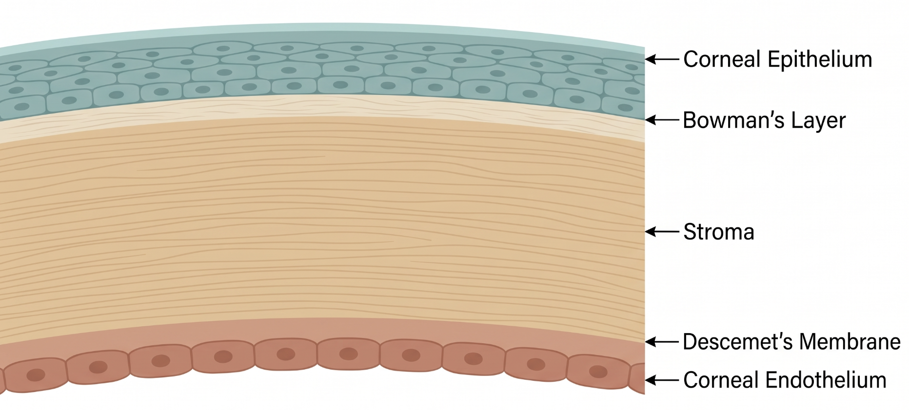
Corneal shape and clarity depend on the structural integrity of the stroma, held together by a highly organized lattice of collagen lamellae cross-linked to one another. Bowman's layer, just beneath the epithelium, provides an additional barrier of structural support. Keratoconus reflects a breakdown of this biomechanical stability.
Keratoconus is a bilateral, typically asymmetric disorder of progressive corneal thinning and steepening (ectasia), producing irregular astigmatism and decreased vision, most often developing in the second and third decades of life.1
Progressive stromal thinning, rupture of Bowman's layer, and ectasia of the central or paracentral cornea are the most common histopathological findings.1 Although traditionally described as noninflammatory, keratoconus has more recently been associated with localized ocular surface inflammation, which may interact with mechanical factors like chronic eye rubbing to drive progressive biomechanical weakening of the stroma.1
Early keratoconus may be asymptomatic or present with mild, progressively worsening blur and glare not fully correctable with spectacles. As disease progresses, patients report frequent glasses-prescription changes, monocular ghosting, and night glare/halos. Acute hydrops — sudden stromal edema from a Descemet membrane break — presents with acute pain, photophobia, and marked vision loss in advanced disease.
Classic exam findings include a scissoring reflex on retinoscopy, Munson's sign, Vogt's striae, and a Fleischer ring.
Differential diagnosis includes pellucid marginal degeneration (inferior thinning, "crab-claw" topographic pattern), post-refractive surgery ectasia, keratoglobus, and contact lens warpage (which can transiently mimic topographic irregularity).
Diagnosis relies primarily on corneal topography and tomography (e.g., Scheimpflug imaging), which detect characteristic anterior and posterior steepening/thinning patterns.1 A single parameter is often insufficient in early/subclinical disease, so pachymetry and higher-order aberration data are increasingly used alongside topography. Machine learning-based algorithms analyzing multiple corneal parameters are an active research area aimed at improving detection of subclinical disease, though no consensus yet exists on the optimal approach.3
Treatment is stepped by severity1:
- Mild disease: spectacles
- Moderate disease: specialty contact lenses (rigid gas-permeable, scleral lenses)
- Progressive disease at any severity: corneal cross-linking (CXL), most effective when performed early
- Severe disease not manageable with scleral lenses: corneal surgery (deep anterior lamellar keratoplasty, or penetrating keratoplasty in advanced/scarred cases)
Sociodemographic disparities in access matter clinically: in one tertiary-center study, Medicaid/Medicare coverage and lower income were associated with more severe disease at presentation and delay in receiving cross-linking.2
- Chronic eye rubbing is a modifiable risk factor — counsel all patients, especially those with allergic eye disease, to stop rubbing
- Corneal cross-linking is most effective when performed early — prompt referral for documented progression matters
- Down syndrome patients have markedly higher keratoconus prevalence and unique diagnostic challenges — maintain a low threshold for screening
- Common pitfall: attributing progressively worsening astigmatism in a young patient to simple refractive error without considering keratoconus
- Common pitfall: missing early/subclinical keratoconus by relying on topography alone without pachymetry or tomography
Keratoconus: From Detection to Treatment Escalation
Clinical Skills for This Topic
Retinoscopy for scissoring reflex
An irregular scissoring reflex on retinoscopy is a classic bedside clue for keratoconus before formal imaging.
Slit-lamp exam for Vogt's striae, Fleischer ring, Munson's sign →
Use cobalt blue light to bring out the Fleischer ring; observe the lower lid contour on downgaze for Munson's sign in advanced disease.
Corneal topography/tomography interpretation (conceptual)
Understand what an asymmetric bowtie or inferior steepening pattern represents, even if you don't operate the device yourself.
Practice Questions
Question 1 of 6
Score: 0/0
What is a major modifiable risk factor for keratoconus progression that should be addressed in every patient?
Saved in this browser.
References
- [1]Santodomingo-Rubido J, et al. Keratoconus: An updated review. Contact Lens & Anterior Eye. 2022.
- [2]Gad RE, et al. Sociodemographic Predictors of Keratoconus (Severity, Treatment, and Progression) in a Tertiary Center in New York City. Cornea. 2024.
- [3]Maile H, et al. Machine Learning Algorithms to Detect Subclinical Keratoconus: Systematic Review. JMIR Medical Informatics. 2021.