
Extraocular Muscle Anatomy & Physiology
Curriculum lesson · updated 2026-07-30
Pending review from section expertOn this page
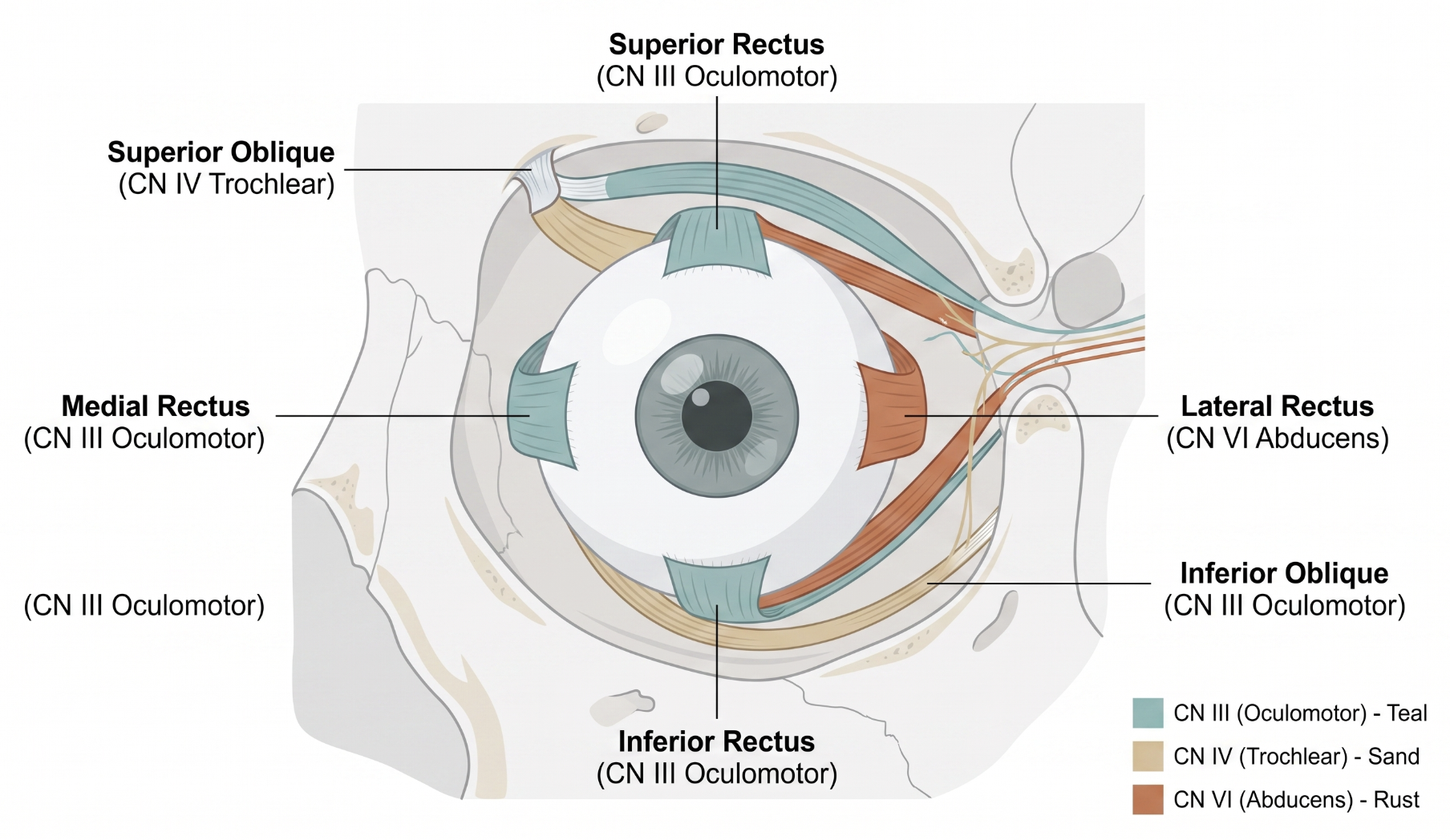
Each eye is moved by six extraocular muscles (EOMs): four rectus muscles (superior, inferior, medial, lateral) originating from the annulus of Zinn at the orbital apex and inserting on the globe anterior to its equator, and two oblique muscles (superior, inferior) with more complex origins/courses inserting posterior to the equator, giving them distinct rotational actions. Cranial nerve III (oculomotor) innervates the superior, inferior, and medial recti and the inferior oblique; cranial nerve IV (trochlear) innervates the superior oblique; cranial nerve VI (abducens) innervates the lateral rectus — the anatomic basis for the motility deficit pattern seen with each cranial neuropathy.
Extraocular muscles are structurally distinct from typical skeletal muscle: most mammalian species, including humans, largely lack the classical proprioceptors (muscle spindles and Golgi tendon organs) that provide position sense in limb muscle.2 Instead, EOMs contain a specialized nerve ending, the palisade ending, whose sensory-versus-motor role was debated for decades; current evidence indicates palisade endings exhibit both sensory and motor characteristics rather than serving a purely sensory position-sensing function as once assumed.2 This means the brain's access to eye position information relies substantially on efference copy and other central mechanisms rather than peripheral proprioceptive feedback in the way limb movement does.2
Coordinated eye movement depends on two classical laws. Sherrington's law of reciprocal innervation describes how, within one eye, contraction of an agonist muscle is accompanied by relaxation of its direct antagonist. Hering's law of equal innervation describes how yoke muscles (paired muscles in each eye moving the eyes together in a given gaze direction) receive equal and simultaneous innervational signals.1 Together, in specific circumstances (such as asymmetrical convergence), these laws can produce isometric antagonist contraction that actually limits eye rotation.1 Passive forces (chiefly natural muscle elasticity) also meaningfully limit rotation: elongation of an antagonist can passively restrict how far an agonist actively rotates the eye, and the system requires dissipative forces to absorb energy between movements, since it is not a perfectly conservative mechanical system.1
Normal binocular vision requires this coordinated motor output be matched by binocular integration in the visual cortex, where neurons combine input from both eyes and become selective for small interocular differences (binocular disparity) to construct stereoscopic depth perception.3 This circuitry develops during a critical period in infancy and childhood, depending on well-aligned, matched input from both eyes during that window.3
- The pattern of ductional limitation on exam can be predicted from the cranial nerve innervating each muscle: CN III (SR, IR, MR, IO), CN IV (SO), CN VI (LR)
- Diagnostic muscle pairs — matching each vertically acting rectus with its corresponding oblique from the other eye — localize a specific underacting or overacting muscle in vertical strabismus1
- Because vertical recti exert stronger vertical action than the obliques, head-tilt testing selectively stresses diagnostic muscle pairs to isolate oblique weakness1
- Disrupted binocular input during the critical period (from strabismus or anisometropia) can prevent normal binocular circuit development, contributing to amblyopia and impaired stereopsis even after later alignment correction3
- Extraocular muscles largely lack classical limb-type proprioceptors, relying instead on palisade endings with mixed sensory/motor character2
- Sherrington's and Hering's laws together explain both normal yoked eye movement and, in some circumstances, apparent paradoxical restriction from antagonist co-contraction1
- Passive muscle elasticity, not just active contraction, meaningfully limits how far the eye can rotate1
- Well-matched binocular input during the critical period is necessary for normal stereopsis circuitry to develop — a key reason early strabismus/amblyopia detection matters3
- Common pitfall: assuming eye movement limitation reflects weakness or restriction alone, without considering passive elastic and reciprocal-innervation contributions1
- Common pitfall: treating extraocular muscle proprioception as equivalent to limb muscle proprioception2
- Common pitfall: assuming animal-model binocular circuitry findings translate directly and completely to humans given acknowledged controversies and gaps3
Six Extraocular Muscles: Innervation & Coordinated Action
Clinical Skills for This Topic
Performing cover/uncover and alternate cover testing →
Detect and characterize misalignment reflecting underlying extraocular muscle or innervational dysfunction.
Testing ductions and versions in the cardinal positions of gaze →
Localize a specific weak or restricted muscle by its predictable pattern of limited movement.
Using head-tilt testing to isolate oblique muscle weakness
Apply diagnostic muscle pair logic to differentiate superior oblique from inferior oblique or rectus muscle involvement in vertical strabismus.
Practice Questions
Question 1 of 6
Score: 0/0
Which cranial nerve innervates the lateral rectus and superior oblique muscles, respectively?
Saved in this browser.
References
- [1]Bicas HEA. A trilogy of the oculomotor system Part II - active, passive, and dissipative forces Part III - diagnostic tests. Arquivos Brasileiros de Oftalmologia. 2026.
- [2]Blumer R, Carrero-Rojas G, Calvo PM, Streicher J, de la Cruz RR, Pastor AM. Proprioceptors in extraocular muscles. Experimental Physiology. 2023.
- [3]Cang J, Fu J, Tanabe S. Neural circuits for binocular vision: Ocular dominance, interocular matching, and disparity selectivity. Frontiers in Neural Circuits. 2023.