
Dry Eye Disease
Curriculum lesson · updated 2026-07-29
Pending review from section expertOn this page
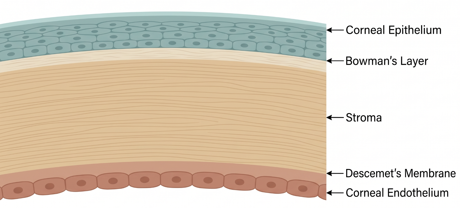
The tear film is a three-layer structure: an outer lipid layer (from the meibomian glands, limiting evaporation), a middle aqueous layer (from the main and accessory lacrimal glands), and an inner mucin layer (from conjunctival goblet cells, allowing the aqueous layer to wet the corneal epithelium). Disruption of any layer can destabilize the whole film.
Dry eye disease (DED) is a common, multifactorial disease of the tear film and ocular surface, defined by TFOS as loss of tear film/ocular surface homeostasis — with tear film instability, hyperosmolarity, ocular surface inflammation and damage, and neurosensory abnormalities as etiological factors — and, importantly, a disease that is always symptomatic.1
A self-perpetuating "vicious cycle" underlies DED: tear film instability leads to hyperosmolarity, which triggers ocular surface inflammation and epithelial damage; that damage further destabilizes the tear film and can cause neurosensory changes, perpetuating the cycle regardless of the initial trigger.2 DED is subclassified into two overlapping mechanistic categories: evaporative (from meibomian gland dysfunction, lid disease, or reduced blink) and aqueous-deficient (from reduced lacrimal tear production, as in Sjögren syndrome).1
Patients report dryness, grittiness/foreign-body sensation, burning, fluctuating vision that improves with blinking, photophobia, and — paradoxically — reflex tearing from surface irritation. Symptoms are often worse later in the day and with prolonged reading or screen use.
Differential diagnosis includes allergic conjunctivitis (itching, papillary reaction), blepharitis/meibomian gland dysfunction (often overlapping and contributory rather than a separate entity), contact lens-related dryness, neurotrophic keratopathy (decreased corneal sensation), medicamentosa from preserved drops, and limbal stem cell deficiency.
Diagnosis follows a structured algorithm: symptom screening with the OSDI-6 questionnaire (cut-off score ≥4), followed by objective testing — non-invasive tear breakup time <10 seconds, tear film hyperosmolarity (≥308 mOsm/L in either eye, or an interocular difference >8 mOsm/L), or corneal/conjunctival staining meeting defined thresholds — any one of which, combined with a positive symptom screen, confirms the diagnosis.1 Subclassification into evaporative, aqueous-deficient, or mixed disease (via meibography, lipid layer assessment, and Schirmer testing or tear meniscus height) then guides targeted therapy.1
Management is stepped by severity:
1. Education, environmental modification, preservative-free artificial tears 2. Lid hygiene and warm compresses for meibomian gland dysfunction; oral omega-3 supplementation is sometimes considered 3. Topical anti-inflammatory therapy (cyclosporine, lifitegrast) or short-course topical corticosteroids for moderate-severe disease 4. Punctal plugs to reduce tear drainage in aqueous-deficient disease 5. Advanced options for refractory disease: autologous serum tears, scleral contact lenses, in-office meibomian gland treatments, or amniotic membrane for severe surface damage
- DED is a disease, not a syndrome, and is by definition always symptomatic per current TFOS consensus
- Reflex tearing/epiphora can be a presenting symptom — don't assume a watery eye rules out dry eye
- Evaporative DED (often from meibomian gland dysfunction) is more common overall than pure aqueous-deficient disease
- Common pitfall: diagnosing DED on symptoms alone without objective confirmatory testing
- Common pitfall: treating all dry eye the same way rather than tailoring therapy to the evaporative versus aqueous-deficient subtype
Diagnosing and Subclassifying Dry Eye Disease
Clinical Skills for This Topic
Tear breakup time (TBUT) measurement
Instill fluorescein and time how long the tear film stays intact before the first dry spot appears; <10 seconds supports the diagnosis.
Slit-lamp evaluation of the lid margin and meibomian glands →
Look for gland dropout, plugged orifices, and abnormal meibum quality to identify an evaporative component.
OSDI-6 symptom screening
Administer the validated questionnaire (cut-off ≥4) as the first step in the diagnostic algorithm, before objective testing.
Practice Questions
Question 1 of 6
Score: 0/0
According to the current TFOS consensus, is dry eye disease considered a syndrome or a disease?
Saved in this browser.
References
- [1]Wolffsohn JS, et al. TFOS DEWS III: Diagnostic Methodology. American Journal of Ophthalmology. 2025.
- [2]Wolffsohn JS, et al. TFOS DEWS II Diagnostic Methodology report. The Ocular Surface. 2017.
- [3]Tsotetsi TP, et al. Dry Eye Disease And The Jenvis Dry Eye Report Diagnosis Tool: A Literature Review with Emphasis On African Populations. Clinical Optometry. 2026.