
Corneal Abrasion & Recurrent Erosion Syndrome
Curriculum lesson · updated 2026-07-29
Pending review from section expertOn this page
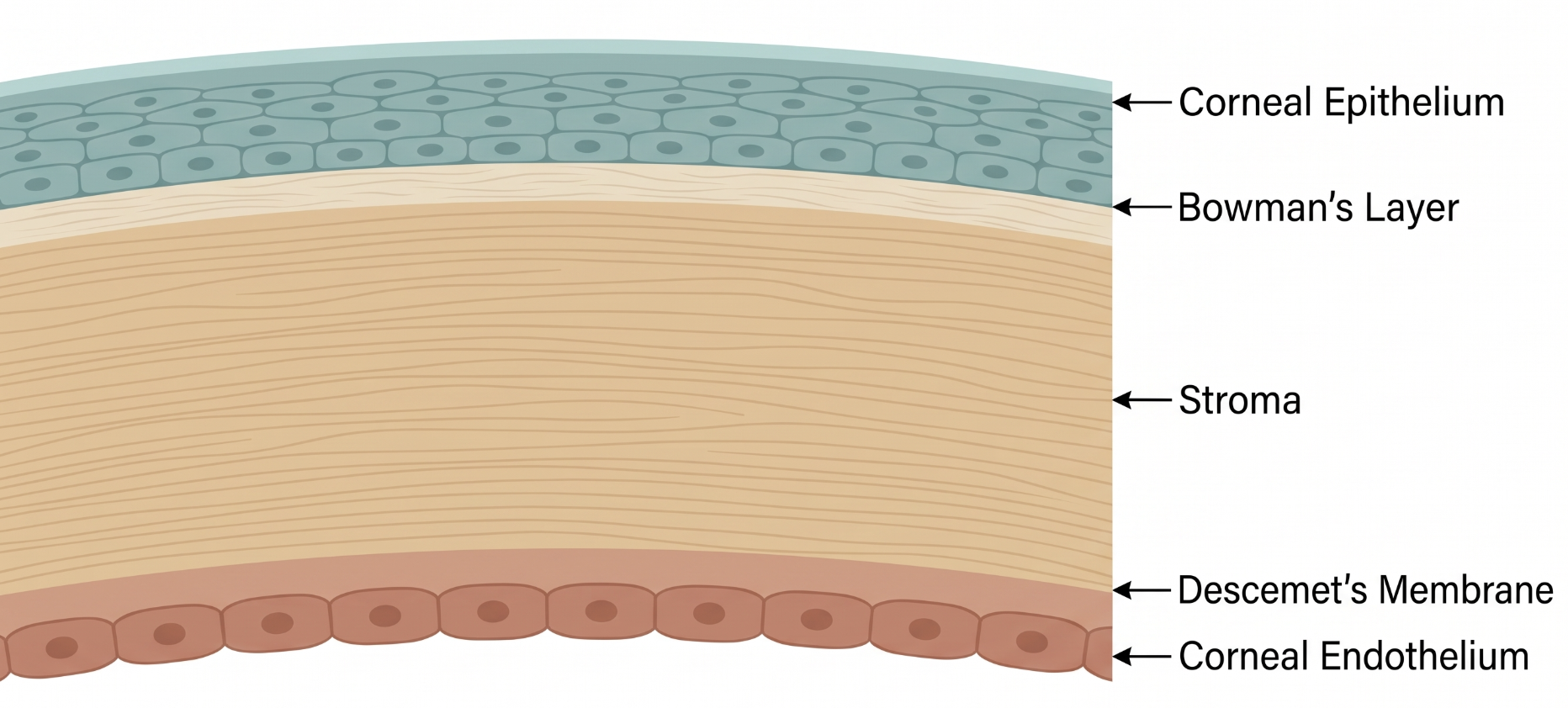
The corneal epithelium is anchored to its basement membrane by hemidesmosomes and anchoring fibrils — an attachment complex that takes weeks to months to fully mature after any disruption, even though the epithelial surface itself typically closes within 24–72 hours. This mismatch between rapid surface healing and slow structural re-attachment is the anatomical basis for recurrent corneal erosion syndrome (RCES).
Corneal abrasion is one of the most common ocular injuries seen in emergency and urgent care settings.1 Most heal uneventfully, but a subset — particularly after fingernail, tree branch, or paper-cut-type injuries — go on to develop RCES, in which the healed epithelium repeatedly breaks down.2
If the epithelium heals with abnormal or incomplete hemidesmosomal adhesion — common after certain injury types or in patients with underlying epithelial basement membrane dystrophy (map-dot-fingerprint dystrophy) — the fragile epithelium can adhere to the tarsal conjunctiva overnight while the lids are closed, then shear off again on eye-opening or blinking upon waking. This is the core mechanism of RCES, explaining its classic waking-hours presentation.
Acute corneal abrasion presents with sudden eye pain, foreign-body sensation, photophobia, tearing, and blepharospasm with a clear trauma history. RCES presents as episodic attacks — classically on waking or eye-opening after sleep — of sudden sharp pain, tearing, redness, and blurred vision, sometimes with a remote history of abrasion months to years earlier.
Differential diagnosis includes herpes simplex keratitis (dendritic staining, decreased corneal sensation — do not mistake for a simple abrasion), a retained subtarsal foreign body (always exclude with lid eversion), contact lens-related keratitis, neurotrophic keratopathy, and epithelial basement membrane dystrophy without any prior trauma history.
Diagnosis is clinical: fluorescein staining under cobalt blue light shows a well-demarcated epithelial defect in acute abrasion. In active RCES, staining may show loose or absent epithelium with a "negative staining" pattern at the leading edge of a healing defect; between episodes, exam may show subtle microcysts or map-dot-fingerprint changes on retroillumination, or can be entirely normal. Lid eversion is mandatory in every case to exclude a retained subtarsal foreign body.
Acute abrasion management is largely supportive: topical antibiotic ointment or drops, cycloplegia for comfort if significant photophobia, and oral analgesia as needed.1 Routine prophylactic patching is generally not recommended, as it does not clearly speed healing and can worsen discomfort or mask complications.1
RCES management is stepped: acute episodes are treated with lubrication, a bandage contact lens, or brief patching for comfort; prophylaxis against recurrence centers on nightly lubricating ointment and/or hypertonic saline (5% sodium chloride), the most common practice change made by ophthalmology in one referral series1; frequent or refractory recurrences may need epithelial debridement, anterior stromal micropuncture, diamond burr polishing, or phototherapeutic keratectomy (PTK) to promote durable epithelial adhesion.
- Always evert the eyelid to exclude a retained subtarsal foreign body in any patient with a corneal abrasion
- Fingernail-induced abrasions, especially from another adult, carry a higher risk of RCES than many other mechanisms
- Hypertonic saline is a simple, common first-line prophylactic measure for RCES
- Common pitfall: missing a dendritic pattern and treating herpes simplex keratitis as a simple abrasion
- Common pitfall: assuming all corneal abrasions require ophthalmology referral, when most heal well with primary/urgent care management alone1
From Acute Abrasion to Recurrent Erosion Syndrome
Clinical Skills for This Topic
Fluorescein staining and cobalt blue light exam
Delineate the size and shape of an epithelial defect, and look for a 'negative staining' pattern in healing recurrent erosions.
Eyelid eversion
Mandatory in every corneal abrasion evaluation to exclude a retained subtarsal foreign body as the ongoing cause of symptoms.
Corneal sensation testing
Check gross corneal sensation to help distinguish a simple traumatic abrasion from herpes simplex keratitis or neurotrophic keratopathy.
Practice Questions
Question 1 of 6
Score: 0/0
Why is lid eversion mandatory in the evaluation of every patient with a corneal abrasion?
Saved in this browser.