
Adenoviral Conjunctivitis (Epidemic Keratoconjunctivitis)
Curriculum lesson · updated 2026-07-29
Pending review from section expertOn this page
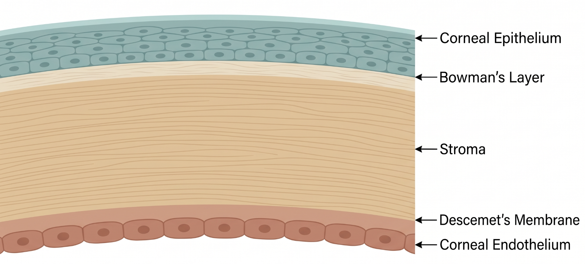
The conjunctiva is a thin, vascularized mucous membrane lining the inner eyelids (palpebral conjunctiva) and the anterior sclera up to the limbus (bulbar conjunctiva), continuous with the corneal epithelium at that border. Its lymphoid tissue and rich vascular supply make it highly reactive to infection and allergy alike, producing the follicular or papillary reactions that help localize a cause.
Adenoviral conjunctivitis is the most common cause of infectious conjunctivitis, ranging from mild follicular conjunctivitis to epidemic keratoconjunctivitis (EKC), in which corneal subepithelial infiltrates develop and can affect vision for months.1 It is highly contagious, spreading via hand-to-eye contact, respiratory droplets, and contaminated surfaces or instruments — including within eye clinics themselves.3
Adenovirus infects conjunctival and corneal epithelial cells, producing an acute follicular inflammatory response. Certain serotypes are particularly associated with the delayed appearance of corneal subepithelial infiltrates, thought to represent a host immune reaction to viral antigen deposited in the anterior stroma rather than ongoing active viral replication at that stage.3 This is why infiltrates can persist long after the patient is no longer contagious.
Three patterns are recognized: epidemic keratoconjunctivitis (watery discharge, marked hyperemia/chemosis, preauricular adenopathy, and — after several days — punctate keratitis progressing to subepithelial infiltrates), pharyngoconjunctival fever (fever, pharyngitis, bilateral conjunctivitis, periauricular nodes, more common in children), and isolated follicular conjunctivitis without corneal or systemic involvement.3 Onset is usually unilateral, spreading to the fellow eye within days.
Overall clinical diagnostic accuracy for viral versus bacterial conjunctivitis is under 50%, so exam findings should be interpreted carefully.3
- Bacterial conjunctivitis: more purulent discharge, papillary rather than follicular reaction, no preauricular node
- Allergic conjunctivitis: itching is prominent, bilateral, papillary reaction
- Herpes simplex keratoconjunctivitis: dendritic epithelial lesions, decreased corneal sensation, usually unilateral
- Chlamydial (inclusion) conjunctivitis: chronic, antibiotic-unresponsive follicular conjunctivitis in sexually active adults
Adenoviral conjunctivitis is primarily a clinical diagnosis.1 Rapid in-office antigen testing is available and can support the diagnosis and reduce unnecessary antibiotic prescribing, though it is not universally used in practice.3 Laboratory viral culture or PCR is reserved for atypical, severe, or outbreak-investigation scenarios.
No antiviral therapy is definitively established for adenoviral conjunctivitis; management is primarily supportive with artificial tears and cool compresses.2 Topical antibiotics are not routinely indicated and should be reserved for suspected bacterial co-infection or high-risk patients such as young children.2
Topical povidone-iodine has emerging evidence suggesting it may reduce viral load and symptom duration, though optimal dosing protocols are still being defined and it is not yet a definitive standard of care.1 Topical corticosteroids are sometimes used for visually significant subepithelial infiltrates or pseudomembranes but can prolong viral shedding, so they should be used cautiously.
Infection control is essential given how contagious adenovirus is: hand hygiene, avoiding shared towels/pillows, instrument disinfection between patients, and counseling patients that they remain contagious for up to about two weeks after symptom onset.3
- Clinical accuracy for distinguishing viral from bacterial conjunctivitis is under 50% — use point-of-care antigen testing when available and don't over-trust gestalt
- Subepithelial infiltrates typically appear 1–3 weeks after onset and can persist for months, well past the contagious period
- Reflexively prescribing topical antibiotics for "pink eye" is a common pitfall when the cause is actually viral
- Pseudomembranes need to be recognized and removed (peeled) to prevent symblepharon formation
- Warn patients about the ~2-week contagious window and clinic-based transmission risk via instruments or multi-dose bottles3
Acute Red Eye: Recognizing Adenoviral Conjunctivitis
Clinical Skills for This Topic
Slit-lamp examination of the conjunctiva and cornea →
Distinguish follicular from papillary conjunctival reaction, and look for pseudomembrane, punctate epithelial keratitis, or subepithelial infiltrates.
Preauricular lymph node palpation
A palpable ipsilateral preauricular node is a classic distinguishing sign favoring viral over bacterial conjunctivitis.
Fluorescein staining of the cornea
Punctate epithelial staining supports viral keratitis; also used to rule out a dendritic pattern suggesting herpes simplex.
Practice Questions
Question 1 of 6
Score: 0/0
A patient presents with acute watery, unilateral red eye with a palpable preauricular lymph node. What is the most likely diagnosis?
Saved in this browser.
References
- [1]Shaikh S, et al. A Comprehensive Review of Adenoviral Conjunctivitis: Exploring the Role of Povidone-iodine in Treatment. International Ophthalmology Clinics. 2025.
- [2]Garcia-Zalisnak D, et al. Adenovirus Ocular Infections: Prevalence, Pathology, Pitfalls, and Practical Pointers. Eye & Contact Lens. 2018.
- [3]Jhanji V, et al. Adenoviral keratoconjunctivitis. Survey of Ophthalmology. 2015.