
Acanthamoeba Keratitis
Curriculum lesson · updated 2026-07-29
Pending review from section expertOn this page
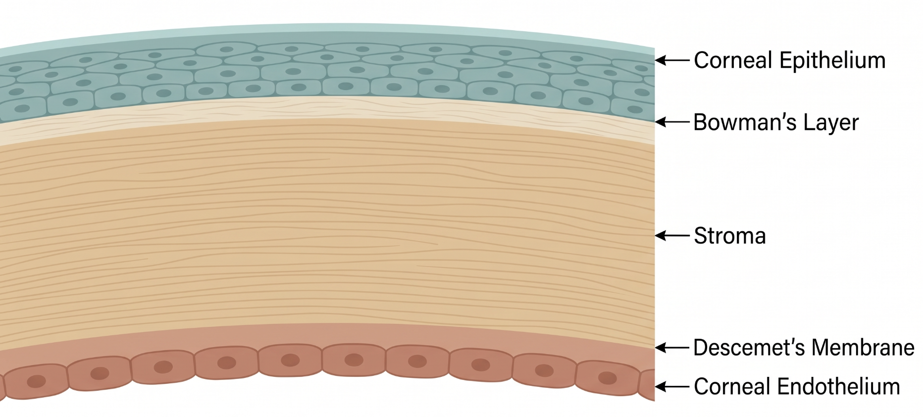
The cornea is one of the most densely innervated tissues in the body, with sensory nerve fibers coursing radially from the limbus toward the center within the stroma and sub-epithelium. This rich innervation is the anatomical basis for the radial perineural infiltrates and disproportionate pain characteristic of Acanthamoeba keratitis (AK), as the organism preferentially tracks along these nerve fibers.
AK is a severe corneal infection caused by Acanthamoeba, a free-living protozoan ubiquitous in soil, dust, and virtually all water sources, disproportionately affecting contact lens wearers.3 It is notorious for diagnostic delay, since early presentation can closely mimic herpes simplex or bacterial keratitis.2
Acanthamoeba trophozoites adhere to and invade the corneal epithelium — facilitated in contact lens wearers by lens-induced epithelial microtrauma — then migrate into the stroma, often tracking along corneal nerves, producing radial perineural infiltrates and severe pain out of proportion to visible findings.3 The organism exists as a motile trophozoite and a highly resistant, dormant double-walled cyst; the cyst form confers substantial drug resistance and allows the organism to persist through treatment and later re-emerge, explaining relapses and the need for prolonged therapy.3
Early AK often mimics herpes simplex or bacterial keratitis. Classic features include severe pain disproportionate to early clinical findings, photophobia, and a waxing-and-waning course despite standard antibacterial/antiviral therapy. Later findings include a ring-shaped stromal infiltrate (specific but late) and radial perineural infiltrates (an earlier, suggestive sign).
Differential diagnosis includes herpes simplex keratitis (most commonly confused early on, given pseudodendrite formation), bacterial keratitis, fungal keratitis, and sterile inflammatory keratitis/neurotrophic keratopathy.
Diagnosis relies on clinical suspicion (contact lens wearer or water/soil exposure, disproportionate pain, refractory course), corneal scraping for culture on non-nutrient agar with an E. coli overlay, and increasingly in vivo confocal microscopy, which can directly visualize cysts non-invasively and has become an important diagnostic tool alongside PCR-based testing and impression cytology.3 Diagnostic delay remains common and is strongly associated with worse outcomes, since antiamoebic treatment is often started only after initial ineffective antibacterial or antiviral therapy has failed.2
Treatment is prolonged, often for many months, centered on topical biguanides (polyhexamethylene biguanide or chlorhexidine) combined with diamidines (propamidine or hexamidine); combination therapy has traditionally been the mainstay, though some evidence suggests biguanide monotherapy at higher concentration may achieve comparable outcomes, an area of ongoing study.1
Topical corticosteroids are controversial: they can reduce inflammatory symptoms but may worsen infection if introduced too early, before adequate antiamoebic control, since they can promote trophozoite proliferation and impair host defenses.1 For advanced or medically refractory disease, therapeutic penetrating keratoplasty may be required, though surgery during active inflammation carries a high risk of graft recurrence.1
- Pain disproportionate to clinical findings in a contact lens wearer, especially with a course refractory to standard therapy, should raise suspicion for AK
- Radial perineural infiltrates are a suggestive early finding; a ring infiltrate is classic but typically appears later
- Diagnostic delay from initially treating for bacterial or herpetic keratitis is strongly linked to worse outcomes
- Common pitfall: mistaking early AK pseudodendrites for herpes simplex keratitis and treating with antivirals alone
- Common pitfall: starting topical corticosteroids before adequate antiamoebic treatment has been established
Recognizing and Treating Acanthamoeba Keratitis
Clinical Skills for This Topic
In vivo confocal microscopy interpretation (conceptual)
Understand that this non-invasive imaging modality can directly visualize Acanthamoeba cysts and has become an important diagnostic tool when available.
Corneal scraping for culture on non-nutrient agar
Requires a specific culture medium (non-nutrient agar with E. coli overlay) distinct from standard bacterial/fungal culture plates.
Contact lens hygiene history-taking
Specifically ask about tap water use for lens cleaning/rinsing, swimming or showering in lenses, and disinfection solution practices.
Practice Questions
Question 1 of 6
Score: 0/0
What clinical clue should raise suspicion for Acanthamoeba keratitis in a contact lens wearer?
Saved in this browser.
References
- [1]Azzopardi M, et al. Advances in medical and surgical management of Acanthamoeba keratitis: a comprehensive review. Clinical Microbiology Reviews. 2026.
- [2]Büchele MLC, et al. Diagnosis and treatment of Acanthamoeba Keratitis: A scoping review demonstrating unfavorable outcomes. Contact Lens & Anterior Eye. 2023.
- [3]Fanselow N, et al. Acanthamoeba Keratitis, Pathology, Diagnosis and Treatment. Pathogens. 2021.